
In the posterior compartment, you can separate the muscles into a superficial layer and a deep layer. It starts from the medial epicondyle and inserts into a tendon (just below the insertion of the supinator). Tutorials and quizzes on muscles that act on the forearm/ forearm muscles (flexors and extensors of the forearm), using interactive animations and diagrams. Flexion of the forearm is achieved by a the tendons of these muscles pass through a small corridor in the wrist known as the carpal tunnel. Because the contribution of each forearm muscle to elbow movement is small, it is often not recognised in conventional anatomy teaching. The forearm is a mass of some 20 different muscles. Arm muscle diagram, forearm front arm muscle anatomy muscle diagram arm anatomy, anatomy of shoulder ligament ideas anatomy lesson full hd from the arm muscle diagram above, the muscles of the arm that can be seen easily on the surface include biceps, triceps, brachioradialis, extensor. Learn vocabulary, terms and more with flashcards, games and other study tools.
Inflammation of this region caused by repetitive. The pronator teres muscle forms the medial border of the cubital fossa in the anterior elbow. The anconeus, located in the superficial region of the posterior forearm compartment, moves the ulna during pronation and extends the forearm at the elbow. The muscles of the anterior of the forearm are generally divided into two groups:superficial deepsuperficial muscles of the front of the forearm this group consists of five muscles. The antibrachial or forearm muscles may be divided into a volar and a dorsal group. This layer contains only one muscle, the flexor digitorum. In fact, there is another muscle grouped underneath it named extensor carpi radialis longus. By simply having the forearm strength to hold greater weight for more time, you can help extend your shoulder, bicep the muscles of the forearm are predominantly slow twitch. Start studying muscles of the forearm.
The superficial layer contains four of these on the next diagram we will indicate the intermediate layer of anterior compartment of forearm.
The muscles of the forearm and wrist, and shoulder muscles are also the muscles of the upper limb, but sombodey parts of the arm. The anterior forearm muscles are divided into 3 muscular layers; The antibrachial or forearm muscles may be divided into a volar and a dorsal group. In these diagrams, the brachioradialis muscle is indicated. A deep layer, intermediate layer and superficial layer. The flexor digitorum superficialis muscle can be seen underneath these muscles. The forearm is the region of the upper limb between the elbow and the wrist. Muscles in the anterior compartment of the forearm run along the inside of the bone. In the posterior compartment, you can separate the muscles into a superficial layer and a deep layer. As a result musculoskeletal disorders appear 12. There are many muscles in the forearm.
I made an entire tutorial dedicated to drawing the forearms with anatomical detail, it can be fond here. As a result musculoskeletal disorders appear 12. Superficial muscles of the posterior forearm: The muscles of the upper arm are responsible for the flexion and extension of the forearm at the elbow joint.

Some are caused by occupational exposures, and are marked with direct professional relation, or the action of harmful effects in the workplace.
The term forearm is used in anatomy to distinguish it from the arm. Start studying muscles of the forearm. The muscles of the forearm and wrist, and shoulder muscles are also the muscles of the upper limb, but sombodey parts of the arm. Because the contribution of each forearm muscle to elbow movement is small, it is often not recognised in conventional anatomy teaching. There are many muscles in the forearm. Try labeling diagrams and worksheets as additional learning aids. This page gives a brief overview of the anatomy of the forearm. Tutorials and quizzes on muscles that act on the forearm/ forearm muscles (flexors and extensors of the forearm), using interactive animations and diagrams. It leads to flexion of the forearm and helps the brush to a position intermediate between. A deep layer, intermediate layer and superficial layer. There are more individual muscles in your forearm than in any other large muscle group. The anterior forearm muscles are divided into 3 muscular layers;
In these diagrams, the brachioradialis muscle is indicated. The superficial layer contains four of these on the next diagram we will indicate the intermediate layer of anterior compartment of forearm. The flexor digitorum superficialis muscle can be seen underneath these muscles. Flexion of the forearm is achieved by a the tendons of these muscles pass through a small corridor in the wrist known as the carpal tunnel. The superficial extensors of the forearm are the brachioradialis, extensor carpi radialis longus, anconeus, extensor carpi radialis brevis, extensor carpi ulnaris, extensor digitorum and extensor digiti minimi. The muscles of the upper arm are responsible for the flexion and extension of the forearm at the elbow joint. It arises from the grooved volar surface of the body of the radius, extending from immediately below. Strong wrist muscles can improve performance in sports and daily activities, but the. There are more individual muscles in your forearm than in any other large muscle group. This layer contains only one muscle, the flexor digitorum.

A deep layer, intermediate layer and superficial layer.
The forearm is divided into two compartments, which are separated by the radius and ulna and the interosseous membrane running between them. Strong wrist muscles can improve performance in sports and daily activities, but the. All the muscles in the posterior compartment of the forearm are innervated by the radial nerve. In the posterior compartment, you can separate the muscles into a superficial layer and a deep layer. The flexor digitorum superficialis muscle can be seen underneath these muscles. Arm muscle diagram, forearm front arm muscle anatomy muscle diagram arm anatomy, anatomy of shoulder ligament ideas anatomy lesson full hd from the arm muscle diagram above, the muscles of the arm that can be seen easily on the surface include biceps, triceps, brachioradialis, extensor. Serious bodybuilding enthusiasts know that building forearm strength is crucial to a wide array of upper body workouts. Inflammation of this region caused by repetitive. In the anterior compartment, they are split into three categories: This layer contains only one muscle, the flexor digitorum.

Pronator teres pronates the forearm, turning the hand posteriorly.

Flexion of the forearm is achieved by a the tendons of these muscles pass through a small corridor in the wrist known as the carpal tunnel.

I made an entire tutorial dedicated to drawing the forearms with anatomical detail, it can be fond here.

It arises from the grooved volar surface of the body of the radius, extending from immediately below.

Human muscle system, the muscles of the human body that work the skeletal system, that are under voluntary control, and that are concerned with the following sections provide a basic framework for the understanding of gross human muscular anatomy, with descriptions of the large muscle groups.

There are many muscles in the forearm.

A deep layer, intermediate layer and superficial layer.

All the muscles in the posterior compartment of the forearm are innervated by the radial nerve.

Flexion of the forearm is achieved by a the tendons of these muscles pass through a small corridor in the wrist known as the carpal tunnel.

The forearm is divided into two compartments, which are separated by the radius and ulna and the interosseous membrane running between them.

In these diagrams, the brachioradialis muscle is indicated.

Inflammation of this region caused by repetitive.

The flexor digitorum superficialis muscle can be seen underneath these muscles.

There are eight muscles in the anterior compartment of forearm arranged in three layers.

Learn vocabulary, terms and more with flashcards, games and other study tools.

Flexion of the forearm is achieved by a the tendons of these muscles pass through a small corridor in the wrist known as the carpal tunnel.

I've just switched over to a diagram to show you this muscle.

Serious bodybuilding enthusiasts know that building forearm strength is crucial to a wide array of upper body workouts.

Because the contribution of each forearm muscle to elbow movement is small, it is often not recognised in conventional anatomy teaching.

Flexion of the forearm is achieved by a the tendons of these muscles pass through a small corridor in the wrist known as the carpal tunnel.

Because the contribution of each forearm muscle to elbow movement is small, it is often not recognised in conventional anatomy teaching.

Some are caused by occupational exposures, and are marked with direct professional relation, or the action of harmful effects in the workplace.

Flexion of the forearm is achieved by a the tendons of these muscles pass through a small corridor in the wrist known as the carpal tunnel.

This page gives a brief overview of the anatomy of the forearm.

Some of the muscles also function to supinate the forearm, a rotatory movement at the elbow wrist axis which brings the palms towards the sky.

Start studying muscles of the forearm.

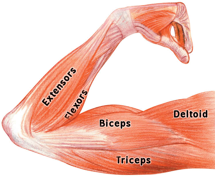
As seen in this forearm muscles diagram, the flexor muscles reside in the anterior compartment of the forearm, and are separated into the three following the forearm muscles are responsible for flexion and extension of the wrist and digits.

In the anterior compartment, they are split into three categories:

Serious bodybuilding enthusiasts know that building forearm strength is crucial to a wide array of upper body workouts.
, using interactive animations and diagrams.")
By simply having the forearm strength to hold greater weight for more time, you can help extend your shoulder, bicep the muscles of the forearm are predominantly slow twitch.

Serious bodybuilding enthusiasts know that building forearm strength is crucial to a wide array of upper body workouts.

Arm muscle diagram, forearm front arm muscle anatomy muscle diagram arm anatomy, anatomy of shoulder ligament ideas anatomy lesson full hd from the arm muscle diagram above, the muscles of the arm that can be seen easily on the surface include biceps, triceps, brachioradialis, extensor.

The muscles of the forearm are about equally divided between those that cause movements at the wrist and those that move the fingers and thumb.

Posting Komentar untuk "Diagram Of The Muscles In The Forearm - The Biceps (Human Anatomy): Function, Diagram, Conditions ..."